The History of Anatomy: Exploring the Human Body Through the Ages
The history of anatomy spans millennia, from ancient Egyptian mummification practices to today's innovative imaging technologies. You'll investigate how Renaissance revolutionaries like da Vinci and Vesalius challenged long-held beliefs, paving the way for scientific breakthroughs. The Age of Enlightenment brought systematic approaches and microscopic examination, while anatomical atlases standardized education. Women's contributions, though often overlooked, have been significant. Modern imaging techniques have revolutionized our understanding of the body's inner workings, and ethical considerations have evolved alongside scientific progress. As you traverse this captivating adventure, you'll uncover how each era has shaped our current knowledge of human anatomy.
Ancient Beginnings
While many ancient civilizations investigated the human body, the Egyptians were among the first to systematically study anatomy. Their mummification practices led to an in-depth understanding of internal organs and their functions. You'll find that Egyptian physicians were skilled in identifying and treating various ailments, thanks to their anatomical knowledge.
In ancient Greece, you'd encounter figures like Hippocrates and Aristotle, who made significant contributions to anatomical comprehension. They challenged prevailing myths and superstitions, laying the groundwork for a more scientific approach to medicine. However, cultural influences and societal perceptions often limited their inquiries, as dissection of human bodies was largely taboo.
The Romans built upon Greek knowledge, with Galen emerging as a prominent figure in anatomical studies. His works, though not always accurate, dominated medical thought for centuries. You'd see his influence persisting well into the Middle Ages.
As you survey ancient anatomy, you'll notice how religious beliefs, philosophical ideas, and societal norms shaped the development of this field. These early efforts, despite their limitations, paved the way for future anatomical revelations.
Renaissance Revolutionaries
Three key figures revolutionized anatomical study during the Renaissance: Leonardo da Vinci, Andreas Vesalius, and William Harvey. Their groundbreaking work laid the foundation for modern anatomy and physiology.
You'll find that da Vinci's detailed anatomical drawings, created through meticulous dissections, were far ahead of his time. He accurately unveiled the human skeleton, muscles, and even the fetus in utero. Though unpublished during his lifetime, his work later influenced medical illustrations.
Vesalius challenged Galenic tradition with his landmark book, "De Humani Corporis Fabrica." You'll see how he corrected numerous anatomical errors and emphasized the importance of direct observation. His accurate depictions of human anatomy transformed medical education and practice.
Harvey's revelation of blood circulation was a pivotal moment in medical history. You'll appreciate how his experiments and calculations proved that the heart pumps blood through a closed system. This revolutionary concept overturned prevailing beliefs and paved the way for understanding human physiology.
These Renaissance revolutionaries' anatomical discoveries and medical innovations propelled the field forward, setting the stage for centuries of scientific advancement in understanding the human body.
The Age of Enlightenment
Building on the Renaissance revolutionaries' pioneering work, the Age of Enlightenment heralded a novel phase of anatomical exploration. During this period, you'd witness a shift towards more systematic and rational approaches to understanding the human body. Philosophical inquiries began to intertwine with scientific methodologies, leading to groundbreaking discoveries and theories.
You'd see anatomists like Giovanni Battista Morgagni pioneering the field of pathological anatomy, linking specific diseases to organ changes. William Harvey's work on blood circulation would revolutionize your understanding of the cardiovascular system. The microscope, refined during this era, would allow you to peer into previously invisible realms of human anatomy.
As you immerse yourself deeper into this age, you'd encounter the rise of experimental physiology, with researchers like Albrecht von Haller conducting meticulous studies on bodily functions. The Enlightenment's emphasis on reason and empiricism would drive anatomists to question long-held beliefs and seek evidence-based explanations. This period would lay the foundation for modern medical practices, transforming your perception of the human body and opening the way for future advancements in anatomy and physiology.
Anatomical Illustrations and Atlases
Anatomical illustrations and atlases played an indispensable role in the advancement of anatomical knowledge. As you immerse yourself in the history of anatomy, you'll uncover that these visual representations revolutionized the way medical professionals and students understood the human body.
In the 16th century, Andreas Vesalius' pioneering work "De Humani Corporis Fabrica" set a new standard for anatomical drawings. His detailed illustrations challenged prevailing misconceptions and paved the way for more accurate depictions of human anatomy. Following Vesalius, other anatomists like Bernhard Siegfried Albinus and William Hunter continued to refine and improve anatomical illustrations.
The 19th century saw a surge in the production of high-quality medical textbook illustrations. Gray's Anatomy, first published in 1858, remains a cornerstone in anatomical education to this day. Its detailed drawings and clear explanations have helped countless medical professionals comprehend complex anatomical structures.
As printing techniques improved, anatomical atlases became more widely available and affordable. These broad collections of anatomical drawings allowed for standardized education and reference materials, profoundly enhancing the study and practice of medicine across the globe.
Microscopy and Cellular Discovery
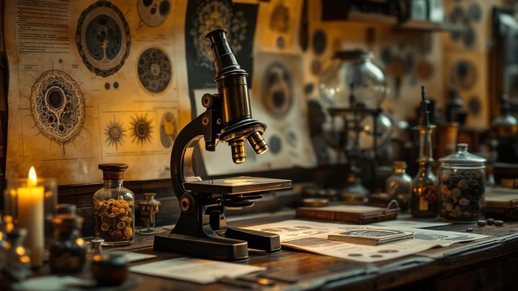
The invention of the microscope in the 17th century ushered in an unprecedented phase of anatomical exploration. You can imagine the excitement of early microscopists as they peered into a hidden world of tiny structures and organisms. Robert Hooke's observations of cork led to the discovery of cells, while Antoni van Leeuwenhoek's simple microscopes revealed bacteria and protozoa.
As microscopy techniques advanced, you'd witness a revolution in our understanding of cell structure and tissue development. The compound microscope allowed for higher magnifications, enabling scientists to study intricate cellular details. In the 19th century, cell theory emerged, proposing that all living things are composed of cells.
This newfound knowledge transformed anatomy and medicine, inspiring awe and wonder:
- Unveiling the building blocks of life
- Revealing the intricate dance of cellular processes
- Discovering the hidden world within our bodies
- Unlocking the secrets of tissue formation
- Bridging the gap between the visible and invisible domains of biology
Microscopy continues to evolve, with electron microscopes and advanced imaging techniques pushing the boundaries of cellular discovery. You're now part of an ongoing expedition to unravel the mysteries of life at its most fundamental level.
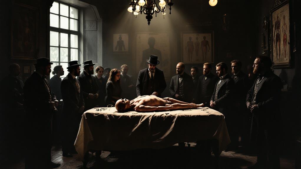
Cadavers and Dissection Practices
While microscopy revolutionized our understanding of life's tiniest components, the study of human anatomy on a larger scale has long relied on a more hands-on approach. Throughout history, cadavers have been essential for medical education and research, allowing students and scientists to investigate the intricacies of the human body firsthand.
You'll find that dissection practices have evolved considerably over time. Ancient Egyptians pioneered embalming techniques, preserving bodies for religious purposes but inadvertently contributing to anatomical knowledge. In medieval Europe, dissections were rare and often conducted in secret due to religious and cultural taboos.
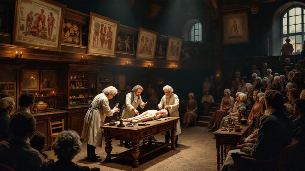
The Renaissance saw a resurgence of interest in human anatomy, with artists and scientists alike studying cadavers to improve their understanding. Anatomical preservation methods advanced, allowing for longer-term examination of specimens. You'd be surprised to learn that public dissections became popular events in the 16th and 17th centuries, drawing crowds enthusiastic to witness the mysteries of the human body uncovered.
Today, modern embalming techniques and plastination have revolutionized cadaver preservation, enabling more detailed and prolonged study. You'll find that these advancements continue to shape our understanding of human anatomy and medical education.
Women in Anatomical Studies

Despite their significant contributions, women's roles in anatomical studies have often been overlooked or minimized throughout history. You'll find that gender biases heavily influenced the field, limiting women's access to education and professional opportunities. However, some exceptional women managed to break through these barriers, making meaningful strides in anatomical research and education.
In the 18th and 19th centuries, women's bodies became subjects of intense medical scrutiny, often leading to medical objectification. This paradoxical situation meant that while women were studied extensively, they were rarely the ones conducting the studies. It wasn't until the late 19th and early 20th centuries that women began to gain more acceptance in medical schools and research institutions.
As you investigate this topic, consider the emotional impact of these historical inequalities:
- Frustration at the lost potential of countless brilliant minds
- Anger at the systemic barriers that held women back
- Admiration for those who persevered against all odds
- Sadness for the women who were objectified in the name of science
- Hope for continued progress in gender equality in medical research
Today, women continue to make significant contributions to anatomical studies, gradually reshaping the field's character.
Modern Imaging Technologies
Moving from historical disparities to technological advancements, we've seen a revolution in anatomical studies through modern imaging technologies. You can now investigate the human body in ways past anatomists could only dream of. Medical imaging techniques like X-rays, CT scans, and MRIs have transformed our understanding of internal structures without the need for invasive procedures.
These technologies allow you to visualize organs, bones, and tissues in incredible detail. You can observe the body's inner workings in real-time, tracking blood flow, brain activity, and even cellular processes. Digital reconstruction models have taken this a step further, enabling you to create 3D representations of anatomical structures. You can manipulate these models, viewing them from any angle and zooming in on specific areas of interest.
Modern imaging has also revolutionized medical diagnoses and treatments. You can now detect abnormalities earlier, plan surgeries with greater precision, and monitor the effectiveness of treatments over time. As these technologies continue to evolve, you'll witness even more impactful advancements in anatomical studies and medical practice.
Ethical Considerations
The study of anatomy has always been intertwined with ethical considerations. As you plunge into the history of anatomical research, you'll encounter numerous ethical dilemmas that have shaped the field. Today, strict guidelines and protocols govern anatomical studies, ensuring respect for human dignity and bodily autonomy.
Informed consent has become a cornerstone of ethical anatomical research. You'll find that modern practices require explicit permission from donors or their families before using bodies for study. Ethical review boards now play a crucial role in overseeing research proposals, ensuring they meet rigorous ethical standards.
The evolution of ethical considerations in anatomy has led to:
- Greater respect for human remains
- Increased transparency in research practices
- Improved protection of vulnerable populations
- Enhanced public trust in scientific institutions
- More inclusive and diverse representation in anatomical studies
As you investigate the ethical terrain of anatomy, you'll discover that these considerations have not only improved the quality of research but also nurtured a deeper appreciation for the human body and the individuals who donate their remains for scientific advancement. The field continues to grapple with new ethical challenges as technology and societal norms evolve.
Future of Anatomical Research
As ethical standards continue to shape anatomical research, state-of-the-art technologies are revolutionizing the field's future. You'll witness pioneering advancements in artificial organs and tissue engineering, which promise to transform medical treatments and our understanding of the human body.
3D bioprinting is at the forefront of this revolution, allowing researchers to create complex tissue structures and even miniature organs for study. You'll see these "organoids" used to test new drugs and therapies, reducing the need for animal testing and accelerating medical breakthroughs.
Virtual and augmented reality technologies are changing how you'll learn anatomy, offering immersive experiences that enhance understanding and retention. These tools will also aid surgeons in planning complex procedures with unparalleled precision.
Advances in imaging techniques, such as high-resolution MRI and micro-CT scans, will provide you with incredibly detailed views of the body's internal structures. This enhanced visualization will lead to new insights about human anatomy and physiology.
As you look to the future, you'll find that anatomical research will increasingly focus on personalized medicine, using genetic information and advanced imaging to tailor treatments to individual patients.
Related posts